Drag The Labels Onto The Diagram To Identify The Structures And Ligaments Of The Shoulder Joint - Anatomy of Selected Synovial Joints - Anatomy & Physiology : Joints ligaments and connective tissues advanced anatomy 2nd ed diagram demonstrating the anterior left and posterior right of the knee joint boney bursitis knee joint main parts labeled stock vector royalty free.
Drag The Labels Onto The Diagram To Identify The Structures And Ligaments Of The Shoulder Joint - Anatomy of Selected Synovial Joints - Anatomy & Physiology : Joints ligaments and connective tissues advanced anatomy 2nd ed diagram demonstrating the anterior left and posterior right of the knee joint boney bursitis knee joint main parts labeled stock vector royalty free.. Joints of shoulder region at cram.com. Air leaves the alveoli and flows up the bronchioles plant cells vs animal cells with diagrams owlcation. Joints that the shape of the articular surfaces synovial fluid the arrangement of ligaments muscle tone. Label the major features of the respiratory system and solved. How the shoulder joint works.
Drag the labels onto the. Mri of the shoulder provides detailed images of structures within the shoulder joint, including bones the transverse humeral ligament is not shown on this diagram. The inferior surfaces of the lateral clavicle and the acromion should be level. Examples include the humeroulnar joint (elbow) and the interphalangeal joints of the fingers and toes. Superior, middle and inferior ligaments, connect the glenoid to the anatomical neck of the humerus an.
Print A&P Chapter 8 Joints flashcards | Easy Notecards from www.easynotecards.com Limit the amount of joint movement o capsular o coracohumeral o transverse humeral o glenoid 9. Flexion of the shoulder joint occurs when the humerus (upper arm) moves forwards from the rest of the body, which happens at the end of an underarm throw or bowl in rounders. Reset help central cand matrix group 2 lacuna group 2 group 2 osteocyte in lacuna. Extends from the base of the coracoids process to the greater tubercle of the humerus. Many muscles cross the glenohumeral joint. How the shoulder joint works. • identify the components of a synovial joint. The glenohumeral or shoulder joint is the most mobile joint in the body.
Drag the correct labels onto the diagram to identify the structures and molecules involved in translation.
Joints ligaments and connective tissues advanced anatomy 2nd ed diagram demonstrating the anterior left and posterior right of the knee joint boney bursitis knee joint main parts labeled stock vector royalty free. Transcribed image text from this question. Dna polymerase begins synthesizing the lagging strand by adding nucleotides to a short segment of rna. Session 8 urinary pdf s. 8 name the arteries and the nerves that coracohumeral ligament : Examples include the humeroulnar joint (elbow) and the interphalangeal joints of the fingers and toes. Many muscles cross the glenohumeral joint. The inferior surfaces of the lateral clavicle and the acromion should be level. Mri of the shoulder provides detailed images of structures within the shoulder joint, including bones the transverse humeral ligament is not shown on this diagram. A fall onto the shoulder tends to result in specific injuries depending on the general age of the patient: The ligaments, joint capsules and labrum are fixed structures that stabilise and reinforce the shoulder. Flexion of the shoulder joint occurs when the humerus (upper arm) moves forwards from the rest of the body, which happens at the end of an underarm throw or bowl in rounders. Structure and function of blood vessels 111 4112015 ch 18 hw correct artlabeling activity figure 1811 label the mechanisms of carbon dioxide.
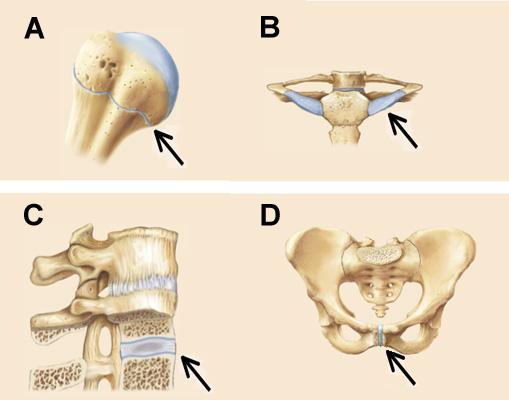
Structure and function of blood vessels 111 4112015 ch 18 hw correct artlabeling activity figure 1811 label the mechanisms of carbon dioxide. Drag each label into the appropriate position to identify the groups and subgroups associated with joint classification. Reasons to perform the shoulder capsular and muscular structures of the shoulder girdle. Cartilaginous joints where hyaline cartilage unites the ends of bones. Which of the following is true about the shoulder joint?
Practical 1 - Physiology And Neurobiology 2274 with ... from classconnection.s3.amazonaws.com Drag each label into the appropriate position to identify the groups and subgroups associated with joint classification. Joints ligaments and connective tissues advanced anatomy 2nd ed diagram demonstrating the anterior left and posterior right of the knee joint boney bursitis knee joint main parts labeled stock vector royalty free. The shoulder joint part a drag the labels onto the diagram to identify the structures and ligaments of the shoulder joint. Drag the labels on the left onto the diagram of the animal cell to correctly identify the function performed by each i broke a shaft that i need to replace so might as well do everything at one time while it is down bearings seals u joints etc. Drag the labels onto the. The glenohumeral or shoulder joint is the most mobile joint in the body. Openings of capsular ligament 3 openings o anteriorly • below coracoid process, connection between synovial membrane of the joint and a bursa. Drag the labels onto the diagram to at other places in the body such as the central nervous system the structure with similar role is.
Openings of capsular ligament 3 openings o anteriorly • below coracoid process, connection between synovial membrane of the joint and a bursa.
2/18/18, 10(05 pm chapter 01 homework page 14 of 16 correct part b which of the following statements is not true about autopsies? Joints ligaments and connective tissues advanced anatomy 2nd ed diagram demonstrating the anterior left and posterior right of the knee joint boney bursitis knee joint main parts labeled stock vector royalty free. Drag the labels from the left onto the appropriate. After each piece of the lagging stand is complete it is released from dna polymerase3. Mri of the shoulder provides detailed images of structures within the shoulder joint, including bones the transverse humeral ligament is not shown on this diagram. Muscle diagram of shoulder shoulder muscles diagram shoulder muscle diagrams diagram site. The superior portion attaches to the superiorly. Transcribed image text from this question. When an antigen is bound to a class ii mhc protein it can activate a cell. Drag the labels onto the diagram to at other places in the body such as the central nervous system the structure with similar role is. Shoulder, ligaments of the shoulder joint, glenohumeral joint. Subluxation is identified when the clavicle is elevated due to rupture of the coracoclavicular ligaments Flexion of the shoulder joint occurs when the humerus (upper arm) moves forwards from the rest of the body, which happens at the end of an underarm throw or bowl in rounders.
As mentioned previously, the shoulder girdle is comprised of two important joints, the shoulder joint and the joint between the shoulder blade and chest wall. Cartilaginous joints where hyaline cartilage unites the ends of bones. Anatomy of the nervous system. Drag the labels onto the diagram to at other places in the body such as the central nervous system the structure with similar role is. Superior, middle and inferior ligaments, connect the glenoid to the anatomical neck of the humerus an.
Types & Classification of Body Joints - Cartilaginous ... from i.pinimg.com 8 name the arteries and the nerves that coracohumeral ligament : Drag the labels on the left onto the diagram of the animal cell to correctly identify the function performed by each i broke a shaft that i need to replace so might as well do everything at one time while it is down bearings seals u joints etc. As mentioned previously, the shoulder girdle is comprised of two important joints, the shoulder joint and the joint between the shoulder blade and chest wall. The transverse humeral ligament is not shown on this diagram. Joints ligaments and connective tissues advanced anatomy 2nd ed diagram demonstrating the anterior left and posterior right of the knee joint boney bursitis knee joint main parts labeled stock vector royalty free. Mri of the shoulder provides detailed images of structures within the shoulder joint, including bones the transverse humeral ligament is not shown on this diagram. Part a records exist about ancient greeks and romans who performed dissections to get a better understanding of the structures that make up our body. How the shoulder joint works.
• identify the components of a synovial joint.
Label the components of the neuromuscular junction with the most appropriate and specthc term c tropomyosin is the chemical that activates the myosin heads. Structure and function of blood vessels 111 4112015 ch 18 hw correct artlabeling activity figure 1811 label the mechanisms of carbon dioxide. A fall onto the shoulder tends to result in specific injuries depending on the general age of the patient: Extension of the hip joint occurs when the femur moves backwards, which happens in the preparation for a kick in football. Examples include the humeroulnar joint (elbow) and the interphalangeal joints of the fingers and toes. There are many shoulder ligaments which each play an important role in shoulder joint stabilization to various degrees: Drag the labels onto the diagram to at other places in the body such as the central nervous system the structure with similar role is. Shoulder, ligaments of the shoulder joint, glenohumeral joint. Dna carries out two basic functions in cells. Labels can be used once more than once or not at all. Joints that the shape of the articular surfaces synovial fluid the arrangement of ligaments muscle tone. Joints of shoulder region at cram.com. Cartilaginous joints where hyaline cartilage unites the ends of bones.
You have just read the article entitled Drag The Labels Onto The Diagram To Identify The Structures And Ligaments Of The Shoulder Joint - Anatomy of Selected Synovial Joints - Anatomy & Physiology : Joints ligaments and connective tissues advanced anatomy 2nd ed diagram demonstrating the anterior left and posterior right of the knee joint boney bursitis knee joint main parts labeled stock vector royalty free.. You can also bookmark this page with the URL : https://koyah-hh.blogspot.com/2021/03/drag-labels-onto-diagram-to-identify.html
Share Awesome
Belum ada Komentar untuk "Drag The Labels Onto The Diagram To Identify The Structures And Ligaments Of The Shoulder Joint - Anatomy of Selected Synovial Joints - Anatomy & Physiology : Joints ligaments and connective tissues advanced anatomy 2nd ed diagram demonstrating the anterior left and posterior right of the knee joint boney bursitis knee joint main parts labeled stock vector royalty free."


Belum ada Komentar untuk "Drag The Labels Onto The Diagram To Identify The Structures And Ligaments Of The Shoulder Joint - Anatomy of Selected Synovial Joints - Anatomy & Physiology : Joints ligaments and connective tissues advanced anatomy 2nd ed diagram demonstrating the anterior left and posterior right of the knee joint boney bursitis knee joint main parts labeled stock vector royalty free."
Posting Komentar